Real-Time Detection
Real-Time Detection
Real-Time Detection
Genetically Encoded Biosensors
LumiRDT® (Real-time Detection Technology) represents LumiSTAR's breakthrough platform of genetically encoded biosensors that transform cellular analysis through superior protein-based indicators.
Unlike traditional chemical fluorescent dyes that suffer from photobleaching, cellular toxicity, and loading variability, LumiRDT® biosensors are genetically encoded within cells, providing unprecedented advantages for drug discovery and biological research.
How It Works
How It Works
How LumiRDT® reagents work
LumiRDT®
reagents work
Our proprietary technology offers long-term detection capabilities extending from
weeks to months compared to hours for conventional chemical probes.
The biosensors demonstrate low toxicity profiles and enable real-time monitoring
of cellular dynamics with superior sensitivity.
Our proprietary technology offers long-term detection capabilities extending from weeks to months compared to hours for conventional chemical probes. The biosensors demonstrate low toxicity profiles and enable real-time monitoring
of cellular dynamics with superior sensitivity.

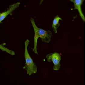
Unlabeled cultured cells are largely transparent, making reliable visualization and morphological analysis of cells and sub‑cellular structures difficult.
LumiRDT engineered viral particles, encoding fluorescent protein reporters, are added to the culture to transduce the cells.
After entry, transduced cells express fluorescent proteins that selectively localize to specific organelles, producing distinct intracellular signals.
These fluorescence patterns are captured by fluorescence microscopy, enabling high‑content, quantitative analysis of cellular architecture and function.

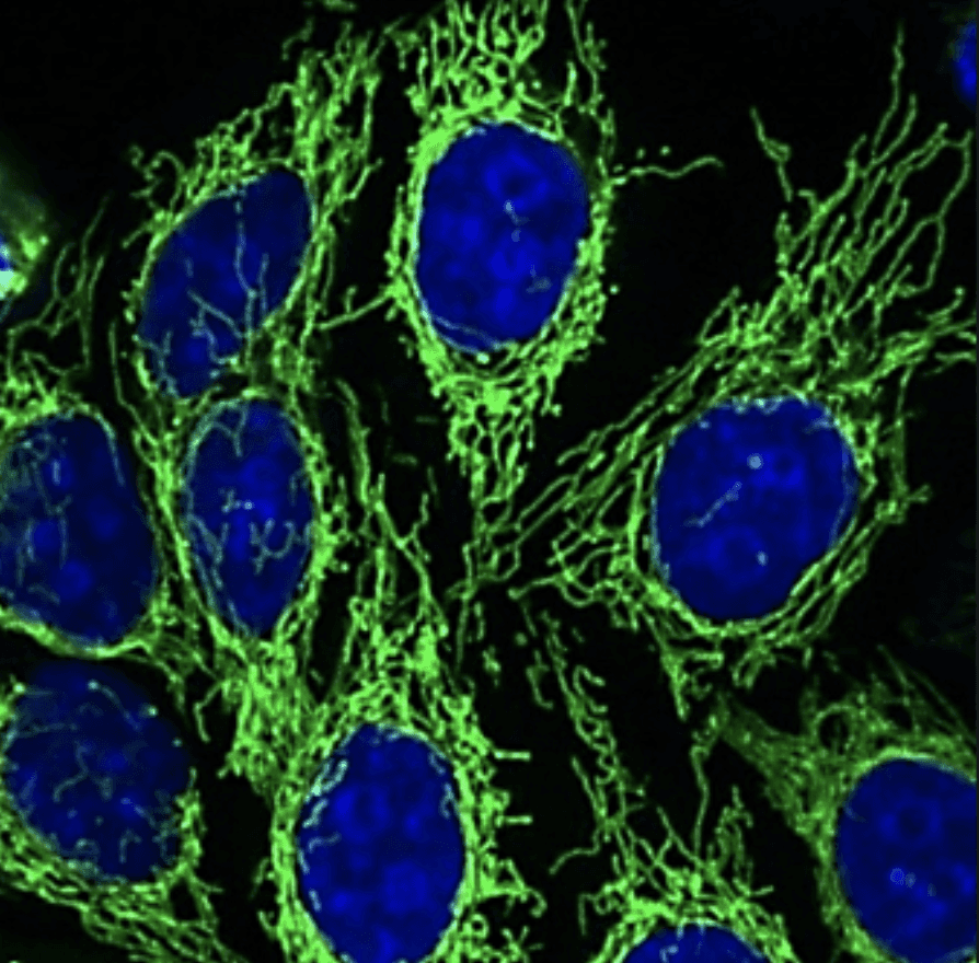

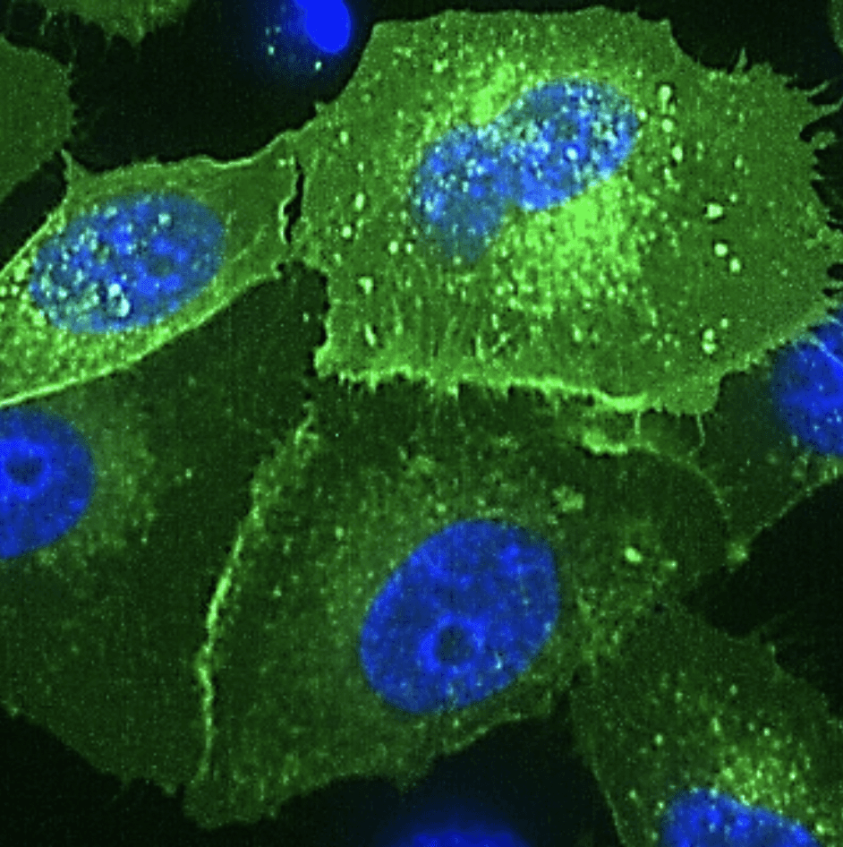

Painter Series
LumiRDT® Painter delivers genetically encoded fluorescent labels in ready-to-use viral reagents. Designed for live-cell imaging, it highlights subcellular structures without fixation or staining, enabling long-term observation. Expressed in dividing or non-dividing cells, LumiRDT® Painter offers multiple colors for simultaneous organelle labeling, enhancing cellular visualization and analysis.
LumiRDT® Painter delivers genetically encoded fluorescent labels in ready-to-use viral reagents. Designed for live-cell imaging, it highlights subcellular structures without fixation or staining, enabling long-term observation. Expressed in dividing or non-dividing cells, LumiRDT® Painter offers multiple colors for simultaneous organelle labeling, enhancing cellular visualization and analysis.
LumiRDT® Painter delivers genetically encoded fluorescent labels in ready-to-use viral reagents. Designed for live-cell imaging, it highlights subcellular structures without fixation or staining, enabling long-term observation. Expressed in dividing or non-dividing cells, LumiRDT® Painter offers multiple colors for simultaneous organelle labeling, enhancing cellular visualization and analysis.
With LumiRDT® Painter you can
With LumiRDT® Painter
you can
• Highlight various organelles with great precision
• Characterize morphological change upon drug treatment
• Conduct chronic studies of cytotoxicity or therapeutic effect with minimal perturbation
• Highlight various organelles with great precision
• Characterize morphological change upon drug treatment
• Conduct chronic studies of cytotoxicity or therapeutic effect with minimal perturbation
Name
Name
Target Organelle
Target Organelle
Color
Color
Data
Data
er.Painter
er.
Painter
Endoplasmic reticulum
Endoplasmic reticulum
Blue
Blue
Green
Green
Red
Red
Far Red
Far Red
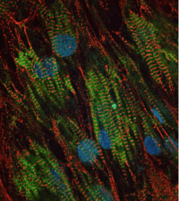


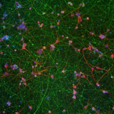
Flash Series
Designed for illuminating calcium ions or reactive oxygen species (ROS) in live cells, LumiRDT® Flash enables real-time and long-term monitoring of organoid health and maturation without fixation or staining. Expressed in dividing or non-dividing cells, LumiRDT® Flash biosensors offer multiple colors and organelle-specific targeting for flexible cellular analysis.
With LumiRDT® Flash you can
• Measure calcium and ROS levels and distribution with great precision
• Monitor and assess organoid maturity over a long period
• Conduct chronic studies of cytotoxicity or therapeutic effect with minimal perturbation
• Measure calcium and ROS levels and distribution with great precision
• Monitor and assess organoid maturity over a long period
• Conduct chronic studies of cytotoxicity or therapeutic effect with minimal perturbation
Name
Name
Localization
Localization
Function
Function
Color
Color
Data
Data
er.Calcium Red
er.
Calcium Red
Endoplasmic reticulum
Endoplasmic reticulum
Ca²⁺ indicator
Ca²⁺ indicator
Red
Red
cyto.Calcium Far Red
cyto.
Calcium Far Red
Cytoplasm
Cytoplasm
Ca²⁺ indicator
Ca²⁺ indicator
Infra Red
Infra Red
FAQ’s
FAQ’s
Frequently Asked Questions
Frequently Asked Questions
/01
What sample types are suitable for LumiRDT®? Does it have specificity?
/02
What are the main differences between LumiRDT® and chemical dyes?
/03
How many samples can one vial of LumiRDT® label?
/04
How should LumiRDT® be used?
/05
Can LumiRDT® be used in animal experiments?
/06
What filter set should be used to image LumiRDT®?
/07
Can LumiRDT® Flash be used for calcium transient experiments?
/08
What should I do if the expression efficiency is low?
/09
How do I label multiple organelles simultaneously?
/10
Can LumiRDT® be customized to suit my application?


Copyright © 2025 LumiSTAR Biotechnology, Inc. All Rights Reserved
Privacy Policy | Terms & Conditions
Copyright © 2025 LumiSTAR Biotechnology, Inc. All Rights Reserved
Privacy Policy | Terms & Conditions
Copyright © 2025 LumiSTAR Biotechnology, Inc. All Rights Reserved
Privacy Policy | Terms & Conditions